Imaging Services for the Enid Area
Medical imaging is the area of medicine that uses X-rays, magnetic waves and ultrasound to obtain detailed images of the inside of the body. Doctors can then use those images to detect and diagnose illnesses and injuries, as well as to help develop treatment plans. The Radiology - Medical Imaging Department at St. Mary's Regional Medical Center offers medical radiology imaging services to residents of Enid, Oklahoma and the surrounding region.
Schedule an Imaging Test at St. Mary's Regional Medical Center
To schedule a radiology exam, please call 580-249-3770.
Types of Imaging Tests Provided by St. Mary's Regional Medical Center
X-ray (Radiography)
X-ray (also called radiography) uses a very small dose of radiation to produce pictures of the inside of the body. X-rays are the most frequently used form of medical imaging and they are also the oldest. They are often used to help see bone fracture, injuries or infections; they are also used to locate foreign objects in soft tissue. In some cases, X-ray exams are used in conjunction with an iodine-based contrast material, which is injected, to help doctors see certain organs, blood vessels or tissue.
Digital Positron Emission Tomography (PET) / Computed Tomography (CT)
A PET scan detects changes in cellular function, specifically how your cells are using nutrients like sugar and oxygen. Since these functional changes take place before physical changes occur, PET can provide information that enables your physician to make an early diagnosis.
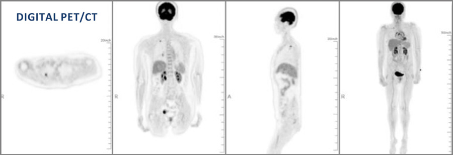
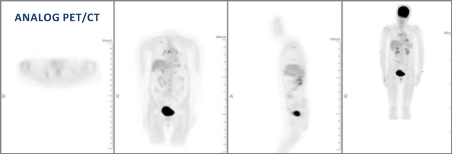
The advantage of CT is its ability to take cross sectional images of your body. These are combined with the information from the PET scan to provide more anatomic details of the metabolic changes in your body.
The PET exam pinpoints metabolic activity in cells and the CT exam provides an anatomical reference. When these two scans are fused together, your physician can view metabolic changes in the proper anatomical context of your body. A PET/CT study not only helps your physician diagnose a problem, it also helps them predict the likely outcome of various therapeutic alternatives, pinpoint the best approach to treatment and monitor your progress.
A PET/CT scan should only take between 10 and 15 minutes. The exam time can vary depending on what we are looking for and what we discover along the way.
Low-Dose CT Lung Cancer Screening
If you smoke, or have quit within the past 15 years, our low-dose CT (LDCT) lung cancer screening test could save your life.
Find out if an LDCT lung cancer screening is right for you >
Magnetic Resonance Imaging (MRI)
Magnetic resonance imaging uses radio waves and a strong magnetic field to create clear, detailed images of internal organs and tissues. Since X-rays are not used, no radiation exposure is involved. Instead, radio waves are directed at the body. This exam takes 30-50 minutes on average and consists of several imaging series. Many studies will require a small intravenous injection of a contrast agent. However, this agent does not contain iodine, an element used in other contrast agents for X-rays or CT scans. MRI is often used to evaluate or diagnose orthopedic (spine, extremity) or neurologic (brain, spinal cord) abnormalities.
Magnetic Resonance Angiography (MRA)
Magnetic Resonance Angiography (MRA) is the use of MRI imaging to study a patient's blood vessels after the injection of a contrast material. Unlike conventional angiography, which is an invasive procedure, MRA is noninvasive other than an injection with a needle to administer the contrast material. This technique can be used to obtain images of arteries in the brain, heart, kidneys, gastrointestinal tract, aorta, neck, chest, limbs and pulmonary system.
Ultrasound
Ultrasound is a type of imaging that uses high-frequency sound waves to produce images of organs and structures inside the body. It is used to view the heart, blood vessels, thyroid, brain, eyes, kidneys, liver, and other organs. It is not good for imaging bones or any tissues that contain air, like the lungs. During pregnancy, doctors use ultrasound to monitor the growth and development of the fetus. Unlike X-rays, ultrasound does not expose patients to radiation.
During an ultrasound test, the patient lies on a table and the technician applies a gel to the skin. This keeps air pockets from forming between the transducer and the skin, which can block ultrasound waves from passing into the body. The transducer sends out sound waves, which bounce off the tissues inside your body. The transducer then captures the waves that bounce back and the ultrasound machine creates images from the sound waves. These images can help doctors determine the location of a structure or abnormality, as well as information about its make up.